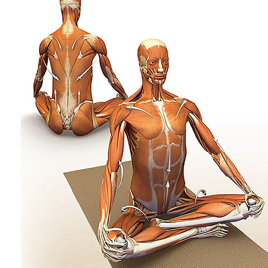
Clinical implications of the Triad of Compression ‘Addressing the relationship between the sacrum, occiput and sphenoid Recently, our community has started moving away from the traditional medical model toward more of a biopsychosocial model of health, writes international lecturer Erin Riley. She says this shift has emerged from the complexity of the illness/wellness dilemma. Manual therapists are at the change, shifting away from a treatment model where the site of the symptoms is the sole focus and are instead looking toward how the entire system is functioning. Manual therapists are looking for root causes of dysfunction, rather than simply treating symptoms. It is no longer plausible to consider the cause of low back painto be localised to the lumbar spine or the cause of neck pain to be localised to the cervical spine. However, we still perhaps have a way to go before we become truly holistic in an approach to bodymindspirit health. CranioSacral Therapy is one of these truly holistic approaches. It considers not only the health and function of our entire interconnected physical body, it considers the mental, emotional and spiritual health of the person. One such global perspective is the consideration and treatment of the relationship between the sacrum, occiput and sphenoid. Dysfunction of these areas and more importantly the reciprocal relationship connecting these dysfunctions has a widespread impact on not just the functioning of the musculoskeletal system, but has a cascade effect on several bodily systems as well as impacting the mental and emotional wellbeing of the client. In CranioSacral Therapy, we call this the Triad of Compression/Depression. What is the Triad of Compression/Depression? The Triad of Compression/Depression was a term coined by American osteopath, Dr John E Upledger, to describe the relationship and impact of compression of three bones simultaneously, the sacrum, occiput and sphenoid. Dr Upledger commonly found that when one of these bones was compressed, it was likely that the other two would also be compressed.1,2,3 Anatomy The relationship between these three bones is not simply an osseous one. The sacrum, occiput and sphenoid are connected through soft tissues and particularly the dural tube and intracranial membrane system. When we begin to understand the intimate and multi- dimensional connections between these structures we can start to conceptualise a framework for comprehending dysfunctions that were previously difficult to solve. Starting at the bottom of this triad, we have the sacrum. The sacrum is a triangle shaped bone that forms the keystone of the pelvis. It articulates with the fifth lumbar vertebrae as well as each ilium at the sacroiliac joint. Normal positioning and functioning of the sacrum is important for both stability and movement. The impact of the Triad of Compression doesn’t end at the level of structural dysfunction. The impact on the central nervous system of a fully compressed sacrum, occiput and sphenoid is that of a ‘pressure cooker’ type situation. Moving further up the system, we come to the occiput. The occiput is located at the base of the skull. Its condyles (just anterior to the foramen magnum) articulate with the superior facet joints of the first cervical vertebrae. Functionally, we need to consider the relationship of the occiput to both the first (C1) and second (C2) cervical vertebrae. The structure of the occipitoatlantal (C0-C1) joint allows for 50 per cent of the cervical spines flexion/extension motion. The relationship between C1 and C2 allows for 50% of the cervical spines rotational movement capacity. 5,6 Also of anatomical importance in the craniocervical region, and a more recent anatomical structure to be written about is the myodural bridge. It has been found that there is a direct relationship between the rectus capitus posterior major and rectus capitus posterior minor musculature and the posterior dural tube via a connective tissue connection called the myodural bridge. This has implications not only in broadcasting tensions up and down the dural tube, but also in broadcasting tension or strain patterns up into the intracranial dura mater. The myodural bridge not only has implications for cervicocephalic pain7,8 but also in sensorimotor control, stabilisation of the spinal cord and monitoring of dural tube tension.8 The third bone in the Triad of Compression is the sphenoid. The sphenoid sits at the back of the orbit of the eye and articulates with many bones of the cranium. The major articulation of importance is between the occiput and the sphenoid at the sphenobasilar junction. Historically, the sphenobasilar junction was considered a symphysis and therefore subject to shearing type motions. Dr Upledger found that the structure of this joint was a synchondrosis and therefore more likely to be impacted on by soft tissue tensions and strain patterns within the intracranial and spinal dura mater. The dura mater is a tough, inelastic, waterproof membrane that lines the underside of the cranial vault and also splits into a second layer that forms the intracranial membrane system. The intracranial membrane system has attachments to the bones of the cranial vault and therefore has a significant influence on compressive and positional dysfunctions of the cranial bones. The dura mater also exits the cranium and forms the spinal dural tube, a mobile sleeve of fascial tissue surrounding the spinal cord. Aside from a strong attachment at the foramen magnum and the second sacral segment and a minor attachment to the posterior bodies of the C2 and/or C3 cervical vertebrae, the dural tube should run relatively unimpeded through the vertebral column.1,4 The dural tube is comprised of three layers of membranes (these layers also extend up into the cranium and intracranial membrane system). It is important for spinal mobility that these layers are able to slide, glide and move independent of each other. This is not only important for mobility but so as to prevent dysfunction being broadcast throughout the system. The dural tube along with the anterior and posterior longitudinal ligaments connect the sacrum and the occiput. Functional relationship It is because of the dural connection that the relationship between the sacrum, occiput and sphenoid is